Skeletal Muscles (A Level only)
1. This question has 4 part(s).
1.1. Figure 1 below shows a cross section through a skeletal muscle.
Figure 1
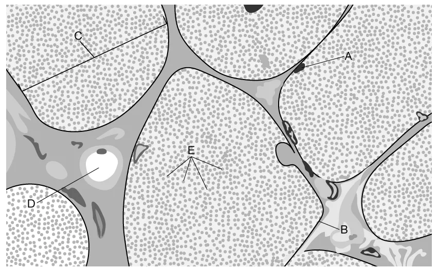
Identify structures A-D labelled on Figure 1.
1.2. The structures labelled E in Figure 1 above are called myofibrils. When myofibrils are viewed in longitudinal section they have a striped, or striated, appearance and are made up of manysarcomeres. Two sarcomeres are shown in Figure 2 below.
Figure 2
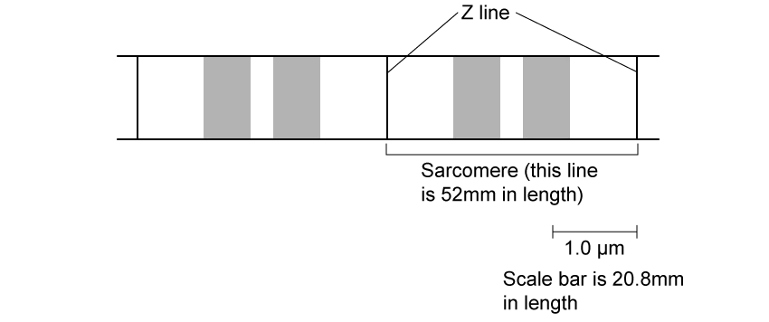
The myofibril is 4.5 cm in length when it is relaxed, while a contracted sarcomere measures 1.9 μm.
Use the scale on Figure 2 to calculate the length of a contracted myofibril (the image measurements are provided for you). Give your answer in mm.
1.3. The striated pattern visible on the sarcomeres is due to the presence of two types of filament within the myofibril. These filaments are known to be involved with muscle contraction, but a relatively recent discovery has shown that a third protein filament, known as titin, is also present. The role played by titin is still being investigated, but one study was carried out in mice that looked at the effects of removing titin from their muscles.
To remove the titin protein, the mice had their muscles modified so that their titin filaments contained a cutting site for a plant enzyme called TEV protease.
Suggest why the plant enzyme TEV was used rather than an animal protease.
1.4. Figure 3 below shows the location of titin within the sarcomere, as well as some of the results gained from the study described in part c) above.
Figure 3
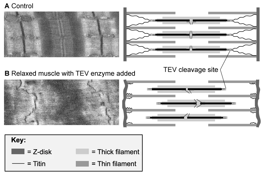
Use Figure 3 and your existing knowledge to suggest 3 possible functions of titin filaments

Ai এর মাধ্যমে

১০ লক্ষ+ প্রশ্ন ডাটাবেজ

প্র্যাকটিস এর মাধ্যমে নিজেকে তৈরি করে ফেলো
উত্তর দিবে তোমার বই থেকে ও তোমার মত করে।

সারা দেশের শিক্ষার্থীদের মধ্যে নিজের অবস্থান যাচাই
No related questions found