9. Gas Exchange
1. This question has 3 part(s).
1.1. Fig. 1 shows two images of lung tissue.
A is a photomicrograph of healthy lung tissue
B is a photomicrograph of lung tissue from a person with emphysema, a chronic obstructive pulmonary disease (COPD)
The images are both at magnification ×40.
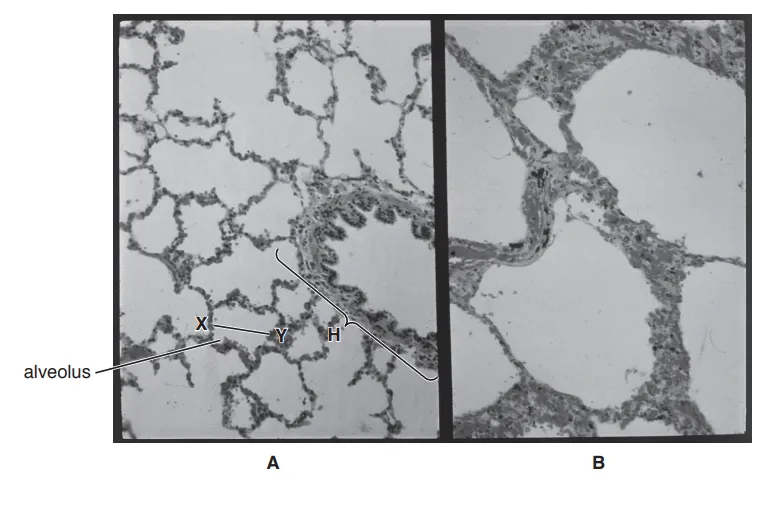
Fig. 1
Name the structure labelled H in Fig. 1A.
1.2. Calculate the actual diameter of the alveolus at X–Y.
Write down the formula and use it to make your calculation.
When measured with a ruler on the page, the image of alveolus X-Y would appear as follows:
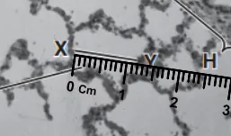
Give your answer to the nearest whole micrometre (μm).
1.3. Gas exchange in the lungs occurs in the alveoli.
Describe the process of gas exchange in healthy lungs and suggest how this compares with gas exchange in the diseased lung shown in Fig. 1B.

Ai এর মাধ্যমে

A database of 1M+ questions

Get yourself ready through practice
Answers from your own book, in your own way.

See where you stand among students across the country
No related questions found